
2026 BRAIN INITIATIVE PHOTO & VIDEO CONTEST
2026 Contest Winners
Congratulations to the 2026 BRAIN Initiative Photo & Video Contest winners!
These inspiring photos and videos highlight the innovation, discovery, and impact of the BRAIN Initiative community. Together, the winning entries reflect the 2026 conference theme, Inventing the Future.
Winning Photos
First Place
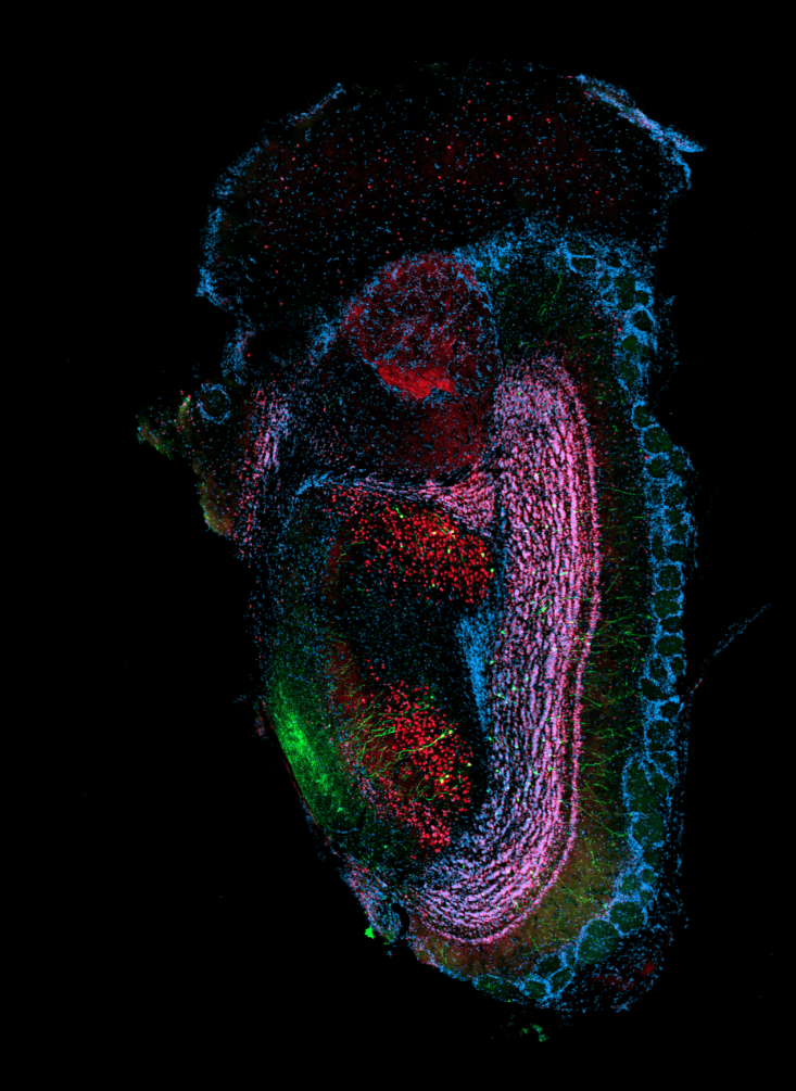
AAV9-mediated GFP expression in the mouse olfactory bulb
by Abigail Nimmo, The Ohio State University
AAV9-GFP was injected into the olfactory bulb of a 7-week-old C57BL/6 mouse to assess transduction efficiency. Brain sections were sectioned at 30 uM. Sections were stained by immunofluorescent-immunohistochemistry (IHC-IF) to visualize transgene expression (GFP, green), neurons (NeuN, red), and nuclei (DAPI, blue). Image acquired at 10x magnification on a Zeiss Axioscan 7 slide scanner.
Second Place
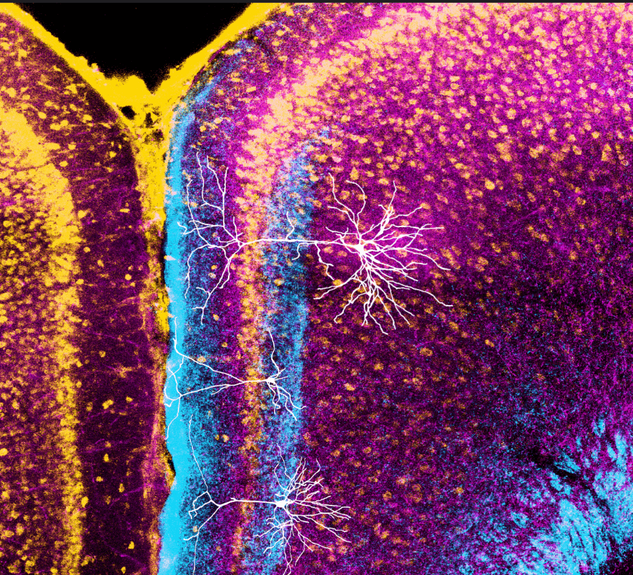
The Little Neuron That Could
By Omar Ahmed, Ph.D., University of Michigan
The retrosplenial cortex is a brain region important for spatial orientation, fear processing, and imagining oneself in the future. The small neuron shown in the middle is unique to this brain region and can fire at high rates. For this reason, the student who identified this neuron liked to call it "the little neuron that could." This neuron receives inputs from head direction cells in the thalamus (shown in blue), unlike its neighboring, more standard neurons (shown above and below it). Thus, this neuron is uniquely positioned to compute spatial orientation information.
Third Place
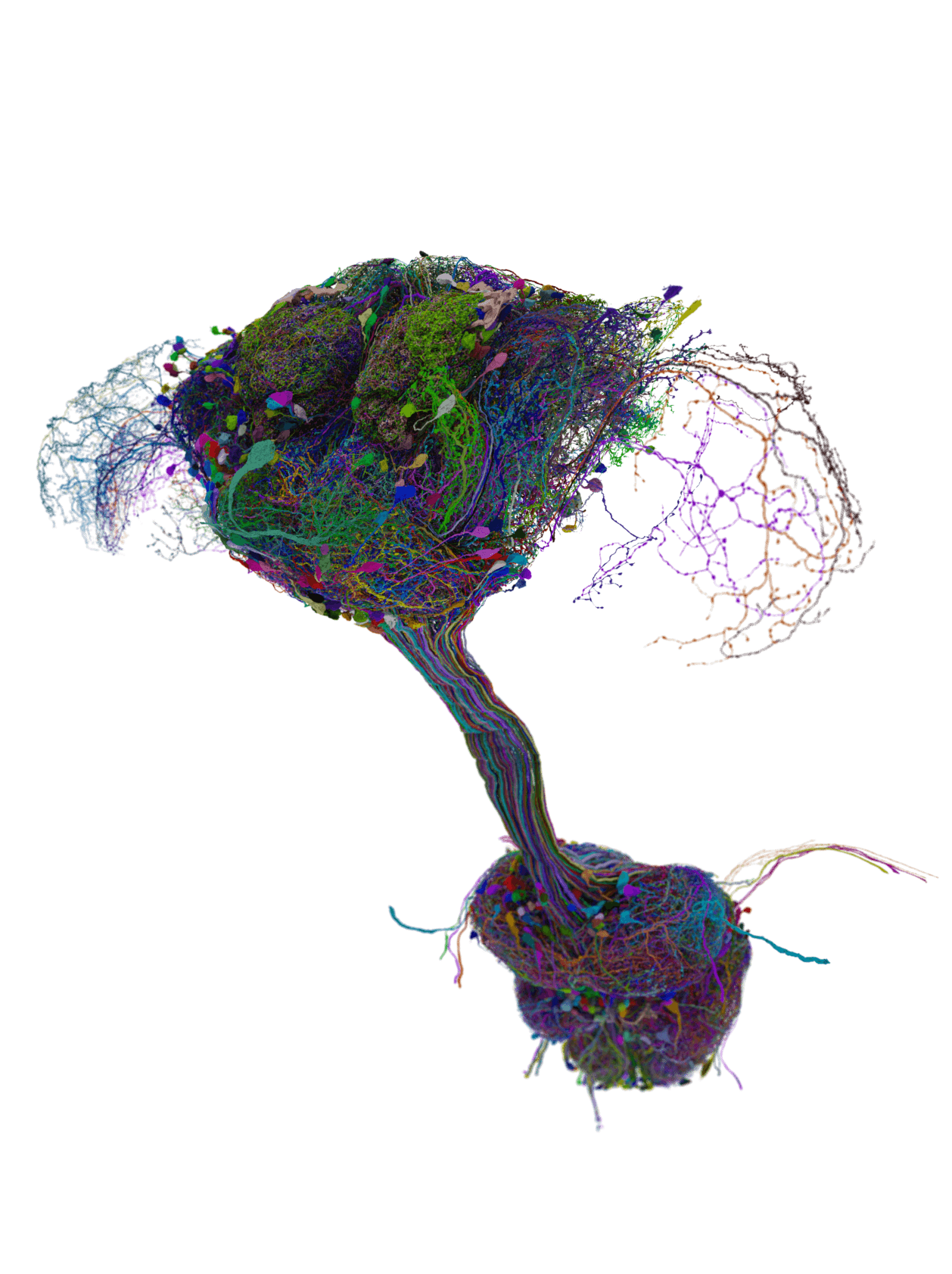
1000 largest neurons in the Drosophila Connectome
by Tyler Sloan, Ph.D., Quorumetrix Studio
This visualization captures the 1,000 largest neurons in the fruit fly central nervous system, offering a comprehensive view by sampling neurons across all functional regions.
Winning Videos
First Place
A Neural Signature of Affective Pain
by Anusha Allawala, Ph.D., University of California, San Francisco and Hazel Huang, University of California, Berkeley
Intracranial EEG recordings reveal changes in the aperiodic exponent across cortical and subcortical networks tracking affective pain symptoms in a patient with refractory chronic pain. Encoding is distributed across both cortical and subcortical regions but lateralized to the right hemisphere, including the right anterior and posterior insula and right midcingulate cortex.
Second Place
Neural enlightenment: restoring cognition through thalamic DBS
by Matthew Hook, Shalini Babu, and Christopher Butson, University of Florida
Ten years ago, the first traumatic brain injury (TBI) patient was implanted with deep brain stimulation (DBS) in thalamus to treat executive function impairment. When asked if she was ready for stimulation, she responded with “light me up!” To date, executive function has been successfully restored in five TBI patients using thalamic DBS. This video demonstrates the modeling of thalamic fibers stimulated during effective therapy in these patients.
Third Place
Multimodal alignments of functional activity, transcriptomics, and projectomics of the same cell
by Lun Wang, Ph.D., Stanford University
Example TRU-FACT (Total Registration Under Functional Activity, Connectivity, and Transcriptomics) alignments of in vivo neural Ca2+ activity and spatial transcriptomic data, using conventional, mesoscope, GRIN microlens-based, or miniature two-photon microscopy in awake mice, together with either HCR-FISH or MERFISH. For multiscale studies of structure-function relationships, TRU-FACT enables routine and scalable multimodal investigations of the same cells by providing a broadly applicable experimental and computational pipeline for image alignment and cell registration.
The intent of this contest was to feature the most colorful and inspiring images that demonstrate today’s advances in neuroscience!
The field of neuroscience has advanced significantly since Santiago Ramón y Cajal, the father of modern neuroscience, created his detailed, hand-drawn illustrations over a century ago. Today, we can capture the intricate beauty and complexity of the brain through modern technology.
As we celebrate the 11th anniversary of the BRAIN Initiative, we continue to uncover the mysteries of the human brain.
Curious about past winners? Check out The Art of the BRAIN: Snapshots from the 2024 Contest or view winners from previous years.